FISH Neutron Imaging
FISH (First Imaging Station Holland), thermal and cold, are two medium-resolution neutron imaging facilities installed at the 2.3 MW research reactor of Delft University of Technology. These instruments serve as a key resource for national and international scientific, education and industry applications. The technique offers non-invasive, in-operando 2D and 3D neutron imaging capabilities, enabling detailed analysis of the bulk of samples at length scales ranging from micrometres to decimetres. Designed to support a diverse range of scientific investigations, the FISH instruments enable the comprehensive studies of materials and structures. They aim to facilitate a wide range of applications, such as internal structure of concrete and engineering materials, inspection of lubricants in engines, internal corrosion inspection, micro-structure of crystallization in steel welds, cracks in composite materials, in-situ study of Li ion batteries, cultural heritage, water uptake in plant roots and soil, rocks etc.
In a typical neutron imaging experiment a sample is irradiated with a polychromatic neutron beam. The sample is placed as close as practically possible to a scintillator-based detector, ensuring maximum resolution. The scintillator screen is made up of a ZnS/6LiF material, which converts neutrons into light. This light is directed towards a camera where a transmission radiograph is recorded. The measurement time for each radiograph can range from 0.5 to 60 seconds, depending on the desired contrast level while ensuring the scintillator screen is not saturated.
To produce final normalized radiograph of a sample, the raw radiographic is normalized against both open beam and dark current measurements. This process corrects for the beam intensity fluctuations, removes the neutron guide structure and background noise, resulting in a more accurate representation of the sample's internal structure.
In 3D computed tomography (CT) measurements, the sample is rotated incrementally, typically from 0 to 360°. At each rotational step, a radiograph is captured. Once all radiographs are collected, the 3D structure of the sample is reconstructed using softwares such as OCTOPUS and MuhRec. For visualization and further analysis, a variety of software tools are available, both commercial and open-source, including AVIZO, VG-Studio, Paraview, Dragonfly, and Fiji (ImageJ). These tools provide powerful capabilities for exploring, quantifying and analyzing the reconstructed 3D data. A typical CT measurement can take several hours, depending on the desired resolution and quality, which can be estimated based on Nyquist-Shannon sampling theorem.
Neutrons offer a unique contrast between elements and isotopes (e. g. Li6 and Li7), combined with high penetration power makes them highly complementary to X-rays. While the X-ray contrast significantly increases for heavier elements, neutrons exhibit strong attenuation for light elements such as hydrogen or boron. Many heavy metal objects appear more transparent to neutrons than to X-rays.
Consequently, neutrons are particularly sensitive to light elements like hydrogen, boron, and lithium, and they penetrate metals (e.g., bronze and steel) more effectively than X-rays. The two imaging instruments are designed in complement with each other, therefore exploring these capabilities further. The thermal FISH situated in the reactor hall utilizes a thermal spectrum, making it ideal for thick metal objects, due to its a high penetration depth albeit at the expense of lower contrast. While, the Cold FISH situated in the experimental hall utilizes the cold spectrum, providing higher contrast, but with lower penetration depth.
Key Features / Advantages for neutron imaging
- Deep Penetration: Neutrons allow the study of bulk and internal structure of materials, and can be combined with complex sample environments like, cryostats, ovens or high pressure equipment
- Sensitivity to Light Elements: Unlike X-rays, neutrons can be highly sensitive to light elements such as H, Li.
- Isotope Differentiation: Neutrons can distinguish between different isotopes of the same element (such as, H and D), for instance allowing to “play” with the contrast by replacing H2O with D2O.
- Magnetic Structure Analysis: Neutrons possess a magnetic moment, enabling them to interact with magnetic moments in materials. This unique property allows us to see internal magnetic features, although this needs a special approach with polarized neutrons as a probe.
Applications
The scientific research conducted using neutron imaging on FISH in Delft is highly diverse. The topics include, but are not limited to
- Non-invasive study of cultural heritage
- Water uptake or transport in various systems like papers, rocks, plants etc.
- Thin glue or lubrication interfaces between metal objects
- Metal corrosion studies within bulk materials
- Crack formation in metal, glass, rocks
- Structure of metal welds, 3D printed metal material, deformation tests
Few examples
1. Neutron Tomography of Van Leeuwenhoek’s Microscopes
The technique of neutron tomography has, after 350 years, enabled a first look inside the iconic single-lens microscopes of Antoni van Leeuwenhoek. Van Leeuwenhoek’s seventeenth-century discovery of ‘animalcules’ marks the birth of microbiology. His skilfully self-produced microscope lenses remained unsurpassed for over 150 years. Neutron tomography now enabled us to reveal the lens types Van Leeuwenhoek used.
Tiemen Cocquyt et al., Neutron tomography of Van Leeuwenhoek’s microscopes.Sci. Adv.7,eabf2402(2021).DOI:10.1126/sciadv.abf2402
2. Chloride-induced corrosion of steel in concrete—insights from bimodal neutron and X-ray microtomography combined with ex-situ microscopy
The steel–concrete interface (SCI) is known to play a major role in corrosion of steel in concrete, but a fundamental understanding is still lacking. One reason is that concrete’s opacity complicates the study of internal processes. The application of bimodal X-ray and neutron microtomography as in-situ imaging techniques to elucidate the mechanism of steel corrosion in concrete. The study demonstrates that the segmentation of the specimen components of relevance—steel, cementitious matrix, aggregates, voids, corrosion products—obtained through bimodal X-ray and neutron imaging is more reliable than that based on the results of each of the two techniques separately.
Angst, U.M., Rossi, E., Boschmann Käthler, C. et al., Mater Struct 57, 56 (2024). https://doi.org/10.1617/s11527-024-02337-7
-
The Fish schematic [1], [2]
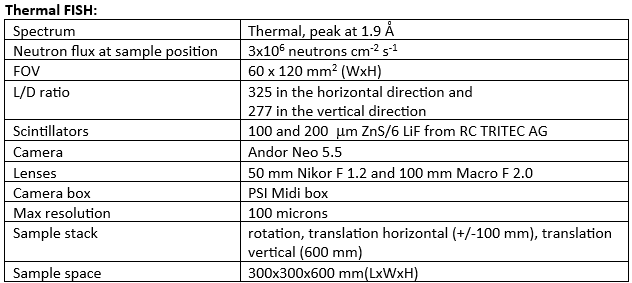
Thermal FISH [1] is installed at the L2 beam port in the reactor hall, with a direct view to the reactor core. The beamline is equipped with a curved stacked neutron guide system, which filters out fast neutrons and gamma radiation. Behind the guide system a shutter is placed, followed by a slit system with a possibility to adjust the height of the beam (standard 40 mm), while the width remains fixed (20 mm). A vacuum tube of 200x200 mm2 minimizes losses, as the beam diverges towards the sample position. The sample stage has computer controlled rotation and translation motors (horizontal and vertical) for scanning the sample, in case the larger field of view (FOV) is required. A scintillator and camera box provides a readout of the radiographs, which are programmed and controlled through a LabVIEW and Python based interface.
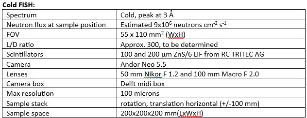
Cold FISH [2] is installed at the R2B beam port in the experimental hall, with a hydrogen (22 K) cold source. Neutrons are transported downstream through a curved neutron guide system that blocks the direct line of sight to the reactor. A main shutter system and a section of straight guides transports the beam to a Li6 aperture that reduces the beam size from 80x20 (HxW) to 30x20 (HxW). A vacuum tube allows the beam to diverge to a FOV of 110 x 55 mm2 (HxW) with minimal losses. The sample stage is equipped with computer controlled rotation and translation motors (only horizontal) for scanning the samples with sizes larger than the FOV of the detector. A scintillator and camera box provides a readout of the radiographs, programmed and controlled through a LabVIEW and Python based interface.
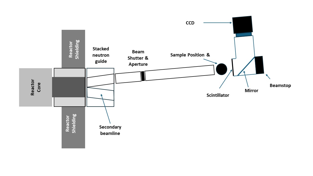
Thermal FISH [1] 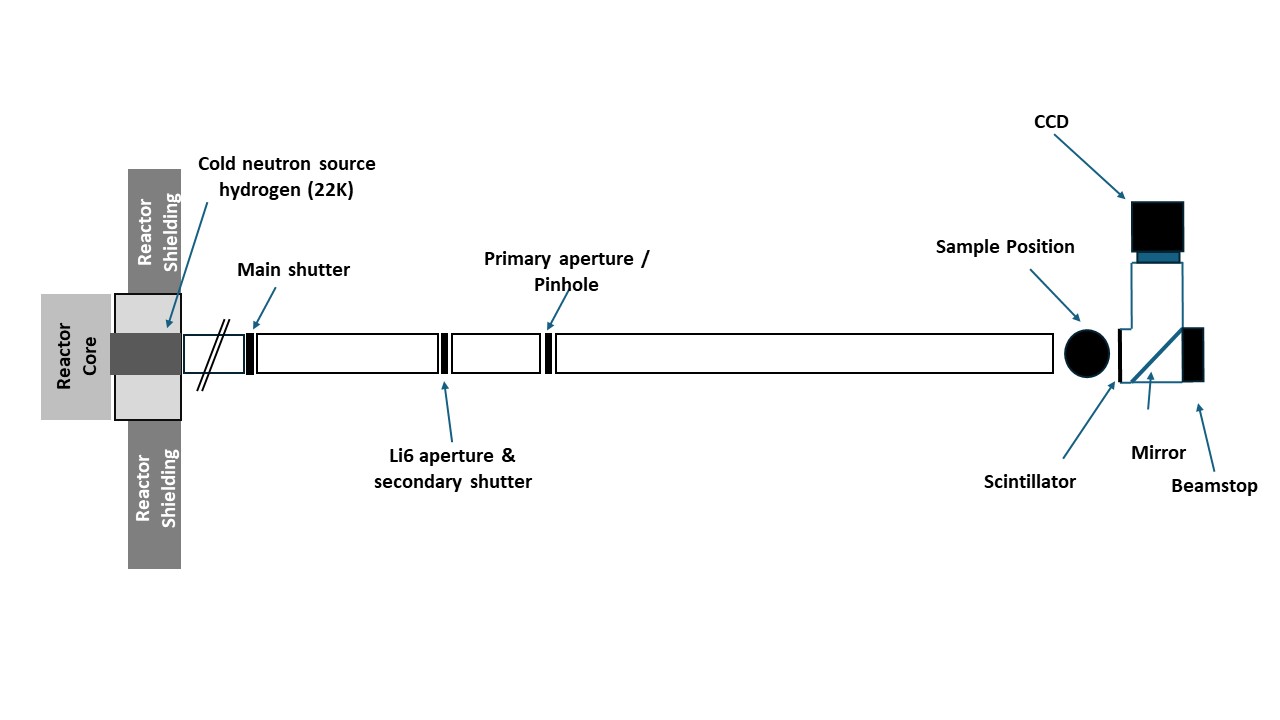
Cold FISH [2] -
Sample size: Samples can range from mm to decimetre size. However, the larger samples are likely to be placed further away from the scintillator detector, hence worsening the resolution.
Safety Considerations
- Radioactivity: If the sample is radioactive, follow all relevant safety protocols for handling and containment.
- Activation: To determine if the sample will be activated due to the exposure to neutrons the composition of the object should be known, if possible.
- Toxicity: For toxic samples, ensure appropriate safety measures need to be taken.
-
There is no specific sample environment for imaging but anything can be discussed. Often bespoke mounting of objects will be arranged.
Please check out the info at the page User Office for Proposal Application, Contact Information and FAQ/QA.
Morphological and microstructural characterization of an ancient Chola bronze statuette by neutron based non-invasive techniques., Cantini, F., Creange, S., Li, Y. et al. Morphological and microstructural characterization of an ancient Chola bronze statuette by neutron-based non-invasive techniques. Archaeol Anthropol Sci 16, 45 (2024).
Change lost: Corrosion of Roman copper alloy coins in changing and variable burial environments, Huisman, L. H., Ackermann, R., Claes, L., van Eijck, L., de Groot, T., Joosten, I., Kemmers, F., Kerkhoven, N., Ngan-Tillard, D. J. M. & More Authors, 2023, In: Journal of Archaeological Science: Reports. 47, 21 p., 103799.
Unravelling the construction of silver filigree spheres from a seventeenth century shipwreck using non-invasive imaging, van der Stok-Nienhuis, J., Beentjes, T., Ngan-Tillard, D., van Eijck, L., Joosten, I. & van Bommel, M. R., 2022, In: Heritage Science. 10, 1, 16 p., 98.
A case study for scientific research prior to conservation of marine metal artefacts, van der Stok-Nienhuis, J., Kuiper, E., Beentjes, T., Joosten, I., van Eijck, L., Zhou, Z. & van Bommel, M., 2021, In: Journal of Archaeological Science: Reports. 37, 102909.
Neutron tomography of Van Leeuwenhoek’s microscopes, Cocquyt, T., Zhou, Z., Plomp, J. & Van Eijck, L., 2021, In: Science Advances. 7, 20, 6 p., eabf2402.
FISH: A thermal neutron imaging station at HOR Delft, Zhou Zhou, Jeroen Plomp, Lambert van Eijck, Peter Vontobel, Ralph P. Harti, Eberhard Lehmann, Catherine Pappas, Journal of Archaeological Science, 20, 369-373 (2018). https://doi.org/10.1016/j.jasrep.2018.05.015.
Gallery
FISH root animation
FISH statue animation
FISH reference sample
FISH cooling channels animation
FISH watch animation
AvLeeuwenhoek microscope

Contact information

dr.ing. J. Plomp (Jeroen)
- +31 (0)15 2787109
- j.plomp@tudelft.nl
-
Room: 2.01.260