Pushing the boundaries of ultrasound
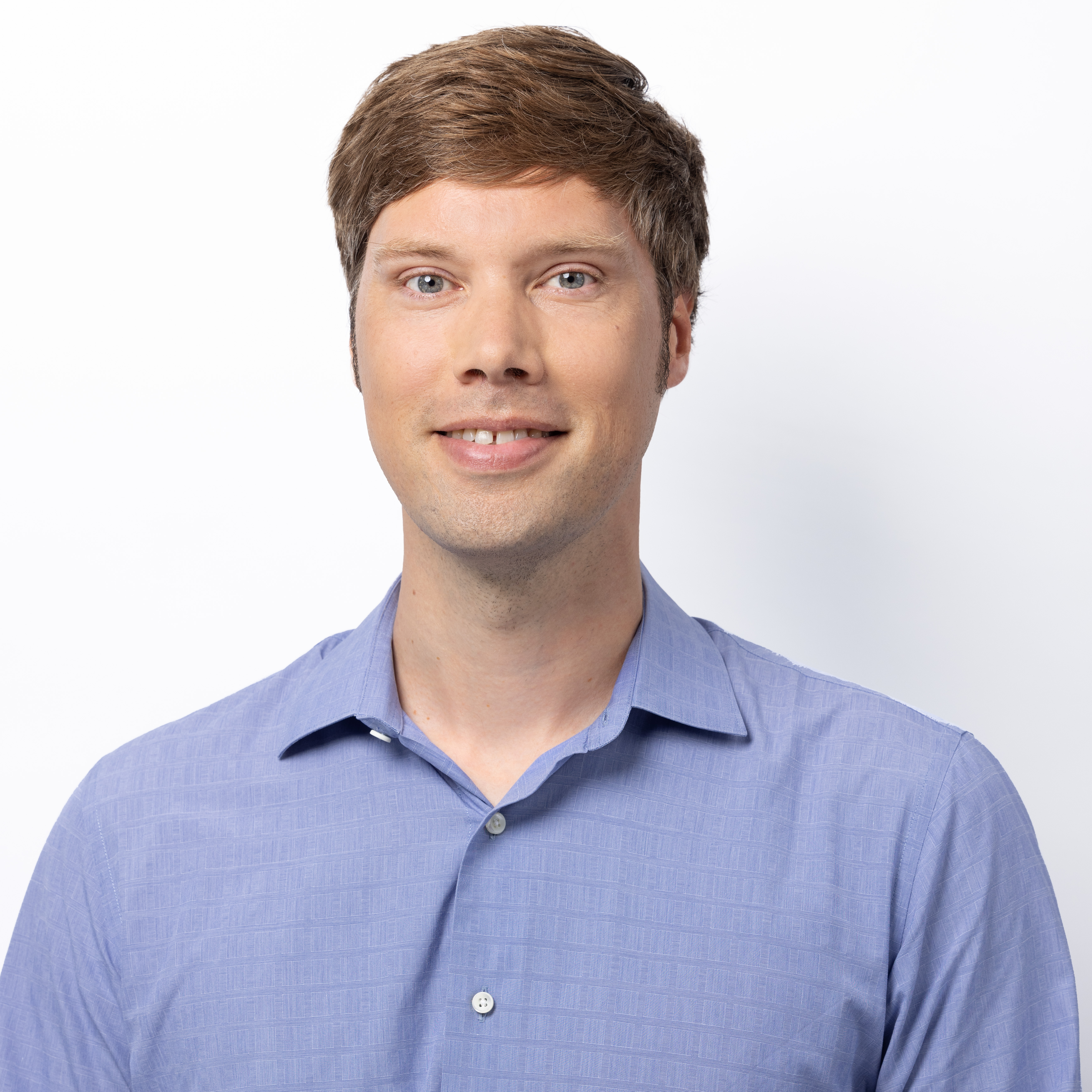
Physicist David Maresca has received a Chan Zuckerberg Initiative Dynamic Imaging grant to develop a next-generation medical ultrasound tool. While state-of-the-art ultrasound imaging, known to most as a baby’s first picture, can show our anatomy and organs, the new tool will be able to zoom in much further, all the way down to the level of the cells in our body. Maresca: “Ultrasound is a safe but also affordable and widespread technology. If we can push the boundaries and make it more sensitive, it will potentially help a lot of people.”

“The goal of my research is to advance the precision of ultrasound imaging down to the cellular level. We want to see how cells behave in the body, in 3D and in real-time,” David Maresca says. Ultrasound imaging reveals the anatomy and motion of internal organs, without causing any harm to the body. “You may be familiar with ultrasound, because for most people, our first picture in life is an ultrasound scan,” says Maresca.
The right treatment
More precise ultrasound can be helpful in diagnosing so-called reperfusion injuries. “Sometimes a blood vessel gets occluded in the brain or in the heart, and all the tissue around that vessel starts to lack of oxygen. That leads to inflammation and this inflammation in turn will trigger immune in tissues,” Maresca explains. “We want to develop a technique that can tell us where the damage occurred, even after the event resolved itself. Was it in the heart or in the brain, which region in the tissue has suffered? That information helps doctors to select the right treatment.”

Noisy proteins
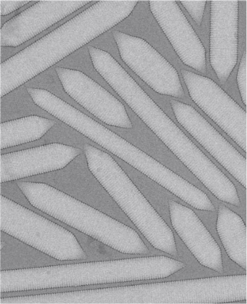
To detect inflammation in the body at the cellular scale, Maresca and his team will use proteins that reflect sound and thus make cells visible under ultrasound – a trick inspired by optics. “In light microscopy, researchers use a fluorescent protein from jellyfish to label cells and follow them in tissues. It was only recently that we found a protein that can do the same in ultrasound. Now, we will further engineer these proteins so that they can sense immune cell activity via changes in acidity in their surroundings, because that’s a marker of inflammation.”
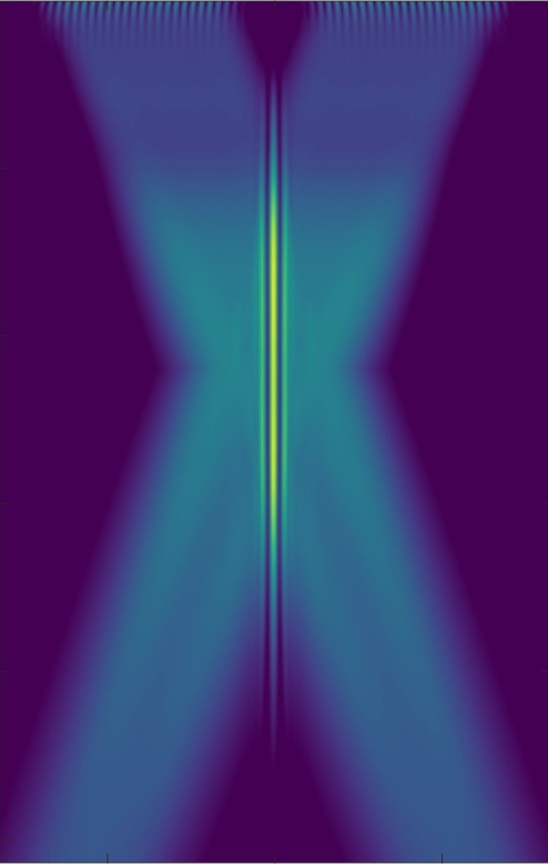
Apart from the noisy proteins, the Delft researchers will be the first to develop sound sheet imaging – an imaging method that already exists for light, but not yet for ultrasound. “You can picture it as a wire that cuts very thin slices of tissue, but non-destructively, it is a harmless process. Once we have taken many of these thin slices in two orthogonal directions, we can reconstruct a 3D volume of tissue, ” Maresca explains.

Memories of heart and brain
Hospitals will be able to use this ultrasound imaging technique to look at a memory of a recent event in the heart or brain, which Maresca illustrates as follows:
“Imagine that a cardiac patient is at home and suddenly feels chest pain. It worries them a bit, because they don’t know exactly what that is. So they go to the hospital to get a scan with ultrasound, even if they have no symptoms anymore. We will be able to tell them whether they shouldn’t worry, because they had a heart burn that can also lead to chest pain, for example. Or we can inform them that something severe did happen in the heart, and what part of the heart suffered precisely. Once the doctor knows that information, they can advise to monitor that patient closely, or channel them through the right standard of care at the hospital. ”
While MRI scanners could give similar information, they are scarce and expensive, making them unsuitable for sorting and channelling patients entering the hospital. “It’s a technique that you use downstream, once you want to confirm a diagnostic,” Maresca says. “Ultrasound is much more versatile in that sense, more flexible.”
The grant is worth 1 million US dollar and allows the hiring of two postdocs, a biotechnician and the purchase of new lab equipment for the next 2.5 years.
The imaging programme of the Chan Zuckerberg Initiative aims to drive the development of new imaging tools that can observe biological processes, as well as the development of robust frameworks to quantify, analyse, and share imaging data and to share methods and tools. To encourage sharing and reuse, imaging sequences developed within this research will be made available through open source licenses wherever possible.

