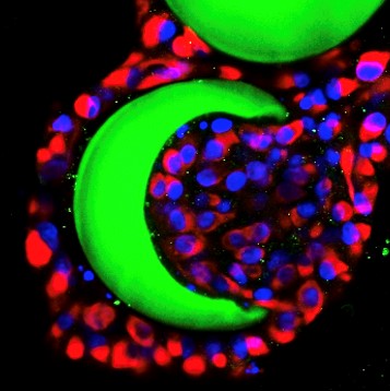
TU Delft and LUMC researchers develop a programmable micro-platform to understand cancer progression and metastasis
Metastasis of tumors is regarded as the largest contributor to cancer-related deaths, but physiologically relevant tumor models for cancer cell metastasis research are still lacking. Two-dimensional (2D) monolayer cell culture (on a flat surface) is traditionally used as in vitro model to investigate tumor behavior, migration, and invasion. Unfortunately, this (2D) monolayer culture does not replicate the complexity of tumor and extracellular microenvironment where invasive cancer cells reside in tumor tissues. Therefore, 3D multicellular systems formed by cancer cells are gaining importance in the field of cancer cell invasion research.
To establish physiologically relevant tumor models, cancer cell spheroids of 3D multicellular aggregates have emerged as a utilizable in vitro model. Although the 2D and 3D tumor models are widely studied, they still exist drawbacks in recapitulating the complex and heterogeneous tumor microenvironment, especially in mimicking precise spatial organization of spheroids and stroma, the dynamic release of cytokines and growth factors, spatiotemporal gradients of functional molecules, and synergy of multiple reactions.
To overcome this limitation, the Boukany group (TUD) closely collaborated with ten Dijke’s lab (LUMC) to develop a new Microbucket-Hydrogel (Mb-H) micro platform for cancer cell invasion. It can be integrated with spatial controlled release of cytokine transforming growth factor-beta (TGFβ) and further programmed with multiple functions to better mimic the complex in vivo microenvironment. Based on this micro platform, multi-cancer cell spheroids were formed, the guiding relationship of single-cell migration and collective cell migration with epithelial-mesenchymal transition (EMT) was demonstrated, and the temporal-spatial controlled manipulation of 3D invasive migration of cancer cells was realized. This programmable and adaptable micro platform addresses the current limitation of constructing an artificial 3D microenvironment by combining microfluidics and soft matter technology to supply an ingenious strategy for 3D cancer cell invasion research. It takes a new step towards mimicking dynamically changing tumor microenvironment and exhibits wide potential applications in cancer research, bio-fabrication, and drug screening to allow for improved personalized treatments of cancer patients. Moreover, it opens a new avenue for recreating 3D in vivo microenvironment and will inspire more future research in this arena.


