443 results

16 June 2021

Structured illumination microscopy with noise-controlled image reconstructions

Structured illumination microscopy with noise-controlled image reconstructions

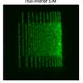
This week a study appeared in Nature Methods on the impact of noise on Structured Illumination Microscopy image reconstructions, and how this knowledge helps to make these reconstructions less sensitive to artefacts as well as to eliminate arbitrary user set parameters. The work was done by Carlas Smith, Kees Hagen, Jacob Hoogenboom, and Sjoerd Stallinga together with PhD-students and collaborators in Erasmus Medical Center and Oxford.

15 June 2021

ZonMw grant for Elizabeth Carroll

ZonMw grant for Elizabeth Carroll

Together with colleagues from other institutions, Elizabeth Carroll has won a grant of up to 750,000 euros in the ZonMw Open Competition. The team will investigate stress in insulin-producing cells.

14 June 2021

Impressive track record: Eric Verschuur conducts EAGE course for 50th time

Impressive track record: Eric Verschuur conducts EAGE course for 50th time

Eric Verschuur was elected in 2006 as the first lecturer for the European Association of Geoscientists and Engineers (EAGE) Education Tour, with 15 lectures in a year throughout Europe, North Africa, Middle East and Russia. Since then he conducts the course 2-3 times a year throughout the whole world.

27 May 2021

Researchers make 3D image with light microscope

Researchers make 3D image with light microscope

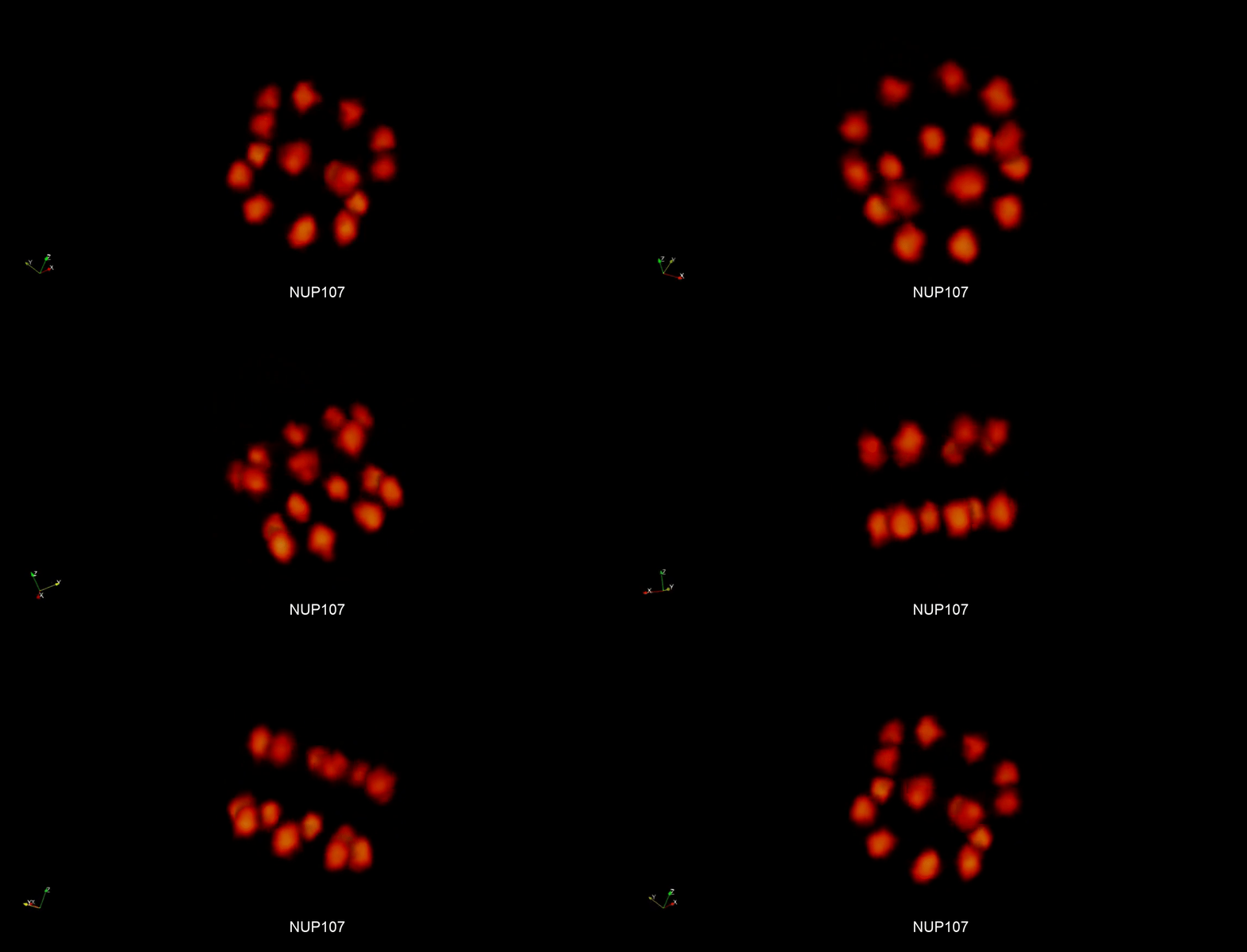
For the first time, Delft researchers have succeeded in making a three-dimensional image of a cellular component using light. The component in question is the nuclear pore complex: tunnels that facilitate traffic to and from the cell nucleus. Studying cell components in 3D can help to determine the cause of various diseases, among other things. The researchers have published their findings in Nature Communications.

27 May 2021

A new tool to understand the brain

A new tool to understand the brain

How does our brain work? An international team of researchers, including lead author Daan Brinks of TU Delft, has taken another step towards answering that question. They have created a new tool that allows them to image electrical signals in brains with an unprecedented combination of precision, resolution, sensitivity, and depth.

26 May 2021

Sobhan Haghparast joined ImPhys as PhD student

Sobhan Haghparast joined ImPhys as PhD student

Sobhan Haghparast joined ImPhys per May 1st. He will work on "Ultra-resolution with visible light" under supervision of Bernd Rieger.

19 May 2021

Tom Haanstra started as technician for the first year physics lab course

Tom Haanstra started as technician for the first year physics lab course

Tom Haanstra recently started working as a technician at ImPhys. His focus will be on the support of the first year physics lab course.

29 April 2021

What's inside the pink box? A nature of science activity for teachers and students

What's inside the pink box? A nature of science activity for teachers and students

Freek Pols published the article: What's inside the pink box? A nature of science activity for teachers and students as part of the dissemination of the Erasmus+ project SPOON. The project aims at professionalizing and supporting science teachers. The article highlights an important but underutilized part of science education: Nature of science. As elaborated in the article, Nature of Science can be taught with simple activities and materials.

28 April 2021

Myrthe Wiersma joined ImPhys as MSc student

Myrthe Wiersma joined ImPhys as MSc student

Myrthe will be working on Ultrasound Localization Microscopy (ULM), supervised by David Maresca and Baptiste Heiles. As a Systems & Control student, she hopes to apply her knowledge on estimation, filtering and optimization to improve the tracking of microbubbles in ULM.

28 April 2021

Delmic: a modern continuation of Delft microscopy heritage

Delmic: a modern continuation of Delft microscopy heritage

Microscopy is inextricably linked to Delft thanks to Antonie van Leeuwenhoek, who was the first to discover cells and microbiological structures with his self-built microscopes in the 17th century. A tradition microscope builder Delmic has been continuing since 2010 with innovative microscope technology. TU Delft Campus interviewed Jacob Hoogenboom of TU Delft who was involved in this innovative company early on and still works closely together with the company.

Agenda

Stay connected

This content is being blocked for you because it contains cookies. Would you like to view this content? By clicking here, you will automatically allow the use of cookies.