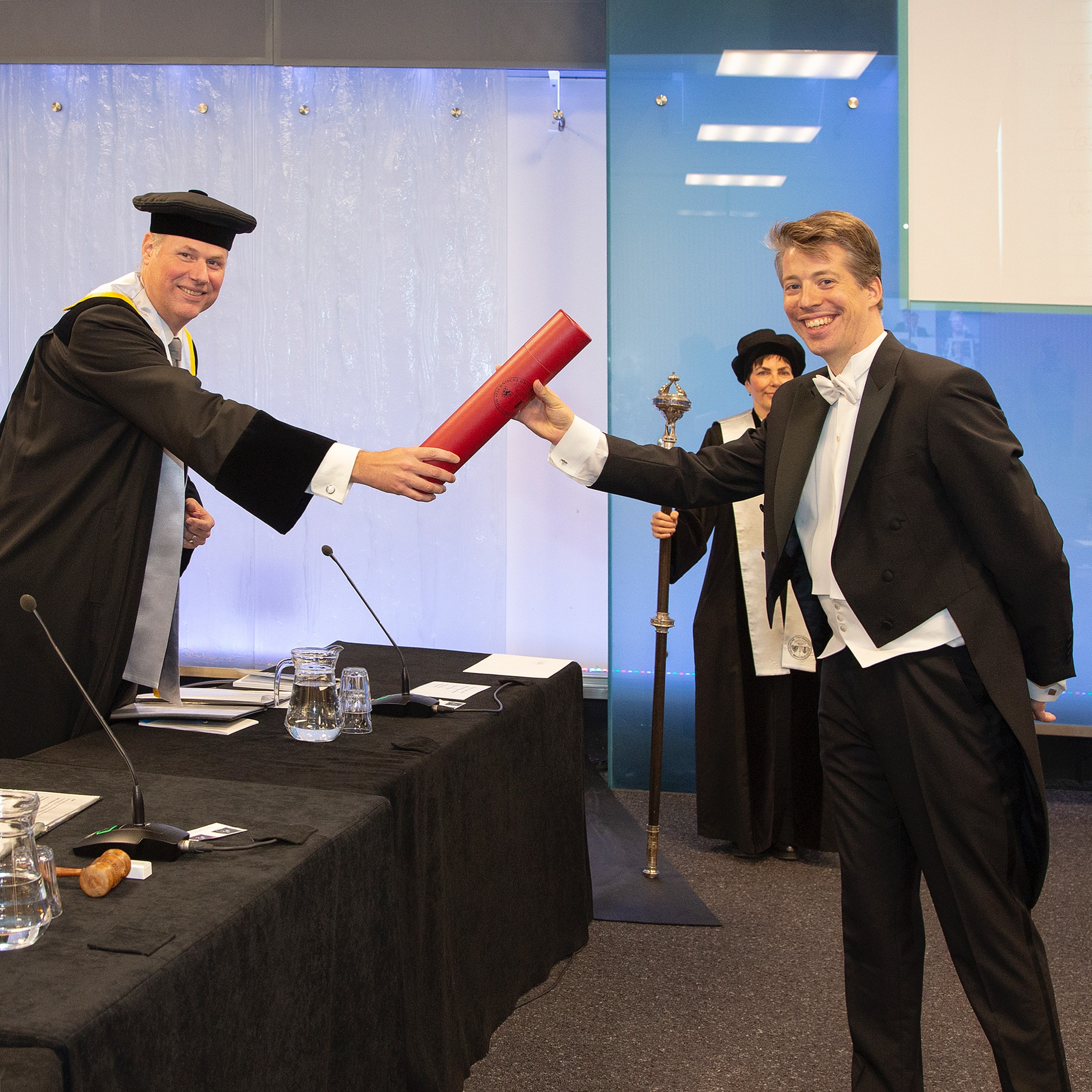
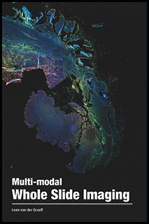
Leon van der Graaff successfully defended his PhD thesis
On September 9th, 2020 Leon van der Graaff successfully defended his PhD thesis on "Multi-modal Whole Slide Imaging".
Abstract
This thesis explores a novel optical architecture for Whole Slide Imaging (WSI). This new architecture allows for multi-focal (3D) image acquisitions in a single scan pass. The multi-focal imaging capability is used to demonstrate 3D phase imaging and 3D imaging of thick tissue sections on a prototype scanner. Further, instrumentation for the extension of WSI to fluorescence imaging is developed: a technologically robust and cost effective method based on LEDs and a highly efficient method based on a multi-line laser illumination source. A WSI system is an optical instrument aimed at creating digital images of bio- logical samples mounted on a microscopy slide at a high throughput. WSI systems image tissues over large fields of view (∼ few cm), in 2D or in 3D (up to hundred layers of μm thickness), and at cellular resolution (∼1 μm). They are applied in high throughput screening in biology, and for novel computer aided medical diagnoses in the field of digital pathology. The optical architecture explored in this thesis is based on a tilted multi-line image sensor concept, originating from Philips. The goal of multi-line image ac- quisition is to enable closed-loop autofocus scanning, but it also allows for multi- focal image acquisitions. The core of this scanner concept lies in a novel design for a multi-line image sensor. The sensor is experimentally characterized for gain and noise, and a system model is developed to find the optimum signal-to-noise ratio (SNR) given the available photo-electron flux. This showed that images with a very high SNR of 292 can be acquired, provided that a sufficiently high photon flux can be realized. Two major issues with the sensor were found in our exper- imental characterization. At high line rates, the sensor showed missing symbols, leading to non-linearities in the read out. Further, the sensor showed high frequent fluctuations in the gain. 3D phase imaging and 3D imaging of thick specimens are two novel contrast modalities based on computational imaging techniques and are enabled by the availability of multi-focal images. For both techniques simplified algorithms were developed compatible with parallel processing at very high speeds. For 3D imaging of thick specimens a deconvolution technique is developed for improving the in- herently low axial contrast. 3D phase imaging is realized by a simplified algorithm for Quantitative Phase Tomography (QPT). QPT imaging is found to be able to im- age the sites labeled for Fluorescence in situ Hybridization (FISH) imaging, and provide additional structural information on unlabeled tissues or tissues stained for immunofluorescence. A system design study is presented showing that the in- plane transfer function has the character of a band-pass spatial frequency filter. The major opportunity for WSI systems to become compatible with fluores- cence imaging is addressed by the development of two imaging modalities. First, a widefield fluorescence WSI system with an LED illumination source is developed and built. A color sequential illumination strategy in combination with multi-band dichroics is used for multi-color imaging using a single monochromatic sensor. The main speed limitation is formed by the exposure time required to capture enough photo-electrons for a decent SNR. Based on the experimental results, a system with 96 Time Delayed Integration (TDI) lines is estimated to achieve a rea- sonable throughput of about 130 kPixel/s. This makes scanning possible of an area of 15 × 15 mm2 in three colors in about 23 min. Second, a novel optical architecture for multi-focal fluorescence image acqui- sitions based on a laser illumination source is proposed and realized in a proto- type. Illumination PSF engineering using diffractive optics is applied to generate a set of parallel scan lines in object space, that span a plane conjugate to a tilted im- age sensor. An important new element in the design is the use of higher order astig- matism to improve the uniformity of peak intensity and line width along the scan lines. Focusing the illumination on the sample provides a very high illumination efficiency and a confocal suppression of background. This optical architecture is projected to ultimately achieve a throughput of several hundreds MPixel/s, which would enable scanning an area of 15 × 15 mm2 in 8 layers in less than a minute. This thesis is concluded with an outlook to opportunities for future research in WSI systems. The challenges and some potential solutions for using a general purpose scientific CMOS (sCMOS) camera for multi-line scanning of a tilted ob- ject plane, and some opportunities for extension of WSI techniques to Light Sheet Microscopy (LSM) and Structured Illumination Microscopy (SIM) are discussed. In summary, this thesis investigates the imaging qualities and extension to computational imaging modalities of a brightfield WSI system and describes two approaches for fluorescence WSI.
https://doi.org/10.4233/uuid:056d0084-880b-438f-9589-e41a108b425a
Contributors
Stallinga, S. (promotor)
van Vliet, L.J. (promotor)

